A New Hope for Type 1 Diabetes?
Could cell transplantation change the future of type 1 diabetes treatment? Discover how engineered islets are showing lasting results now!
A novel therapeutic strategy has demonstrated success in reversing type 1 diabetes in preclinical studies. Insulin-producing pancreatic islet cells, when transplanted alongside specially engineered cells that promote blood vessel formation, were found to restore healthy glucose regulation.
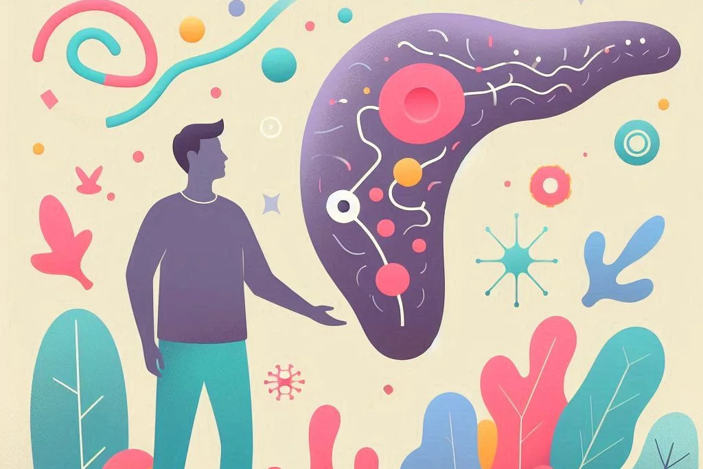
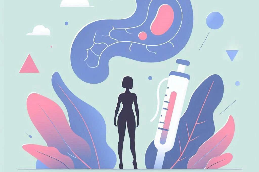
The pancreatic islet is uniquely responsible for insulin secretion in response to increased blood sugar levels. In cases of type 1 diabetes, this function is impaired due to an autoimmune response that targets and gradually eliminates these insulin-producing cells. Advances in islet transplantation have offered a promising avenue for restoring insulin production and improving metabolic health.
One of the main obstacles has been the recreation of the vascularized environment essential to islet cell function. Recent research conducted at Weill Cornell Medicine has addressed this issue by combining islet transplantation with vascular-supportive cells. In experimental models, this method resulted in the complete reversal of diabetic symptoms.
With continued research and validation, this approach holds the potential to transform the management of a condition that has remained without a cure.

A foundational step has been taken toward establishing subcutaneous islet transplantation as a potentially safer and more lasting treatment for type 1 diabetes, as noted by the study’s lead author, Ge Li, PhD, from the Department of Medicine at Weill Cornell Medicine.
The current standard for islet transplantation typically involves transferring donor islets into the hepatic portal vein, often through a minimally invasive procedure targeting the liver. Within this environment, islets settle into small blood vessels known as sinusoids, gradually integrating with surrounding tissue as new vasculature forms. However, a significant portion of islets may be compromised during this period due to inflammation, limited oxygen supply, or immune system activity. Long-term administration of immunosuppressive medications is generally required to minimize rejection.
In pursuit of a less invasive and more sustainable solution, researchers sought to identify an alternative implantation site with improved accessibility and enhanced survival potential for donor islets. To this end, human endothelial cells—normally responsible for lining blood vessels—were bioengineered into reprogrammed vascular endothelial cells, or R-VECs. These were initially assessed using a microfluidic platform, where they successfully organized into functional vascular networks capable of conducting human blood.
When introduced alongside human islets, the R-VECs facilitated a supportive vascular environment. The islets became embedded within these newly formed vessels, which both surrounded and infiltrated the clusters. This engineered blood supply enabled the islets to perform effectively, including insulin release in response to glucose presence.
Such developments reflect a significant stride toward expanding the therapeutic potential of islet transplantation, with emphasis on accessibility, durability, and functional integration.
In a significant advancement, human islets and R-VECs were co-transplanted beneath the skin of diabetic mice. As observed in earlier laboratory experiments, the transplanted components successfully formed a vascularized islet network. This network enabled the production of human insulin, resulting in normalized blood glucose levels for over 20 weeks. The extended duration of glucose control in the mice strongly indicated the long-term viability of the islet/R-VEC graft. In contrast, mice that received only islets—without the vascular support—exhibited markedly reduced insulin output and lacked proper glucose responsiveness.
“Remarkably, it was observed that R-VECs did adapt when co-transplanted with islets, supporting the islets with a rich mesh of new vessels and even taking on the gene activity ‘signature’ of natural islet endothelial cells,” noted David Redmond, assistant professor of computational biology research in medicine at Weill Cornell Medicine.

Ongoing efforts are focused on continuing preclinical evaluations to ensure both safety and sustained effectiveness of the implant.
“Ultimately, the potential of surgical implantation of these vascularized islets needs to be examined for their safety and efficacy in additional preclinical models,” stated Dr. Rebecca Craig-Schapiro, associate professor of surgery at Weill Cornell.
Looking ahead, hopes remain high for the introduction of this novel transplantation method as a treatment option for type 1 diabetes in the near future.
“Translation of this technology to treat patients with type 1 diabetes will require circumventing numerous hurdles, including scaling up sufficient numbers of vascularized islets, and devising approaches to avoid immunosuppression.”
These findings offer a compelling step forward in regenerative strategies aimed at achieving long-term metabolic health without reliance on external insulin administration.
https://doi.org/10.1126/sciadv.adq5302
Summary
Tissue-specific endothelial cells (ECs) are critical for the homeostasis of pancreatic islets and most other tissues. In vitro recapitulation of islet biology and therapeutic islet transplantation both require adequate vascularization, which remains a challenge. Using human reprogrammed vascular ECs (R-VECs), human islets were functionally vascularized in vitro, demonstrating responsive, dynamic glucose-stimulated insulin secretion and Ca2+ influx. Subcutaneous transplantation of islets with R-VECs reversed hyperglycemia in diabetic mice, with high levels of human insulin detected within recipient serum and relapses of hyperglycemia following graft removal. Examination of retrieved grafts demonstrated that engrafted human islets were mainly vascularized by the cotransplanted R-VECs, which had anastomosed with the host microcirculation. Notably, single-cell RNA-sequencing revealed that R-VECs, when cocultured with islets, acquired islet EC-specific characteristics. Together, R-VECs establish an adaptable vascular niche that supports islet homeostasis both in vitro and in vivo.